 Return to Methods List |
Methods in Neuroscience Manduca sexta Segmental Ganglia - Recording p. 3 |
|
Some recording basics Click HERE for a 2 minute video (18.2MB) |
|
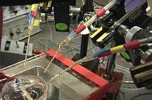 Here you see the recording dish and the three suction recording electrodes. |
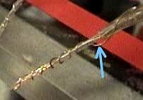 Wrapped around each suction recording electrode is a ground wire (blue arrow). |
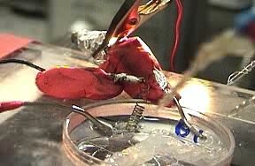 Here is a close-up view of the recording dish. |
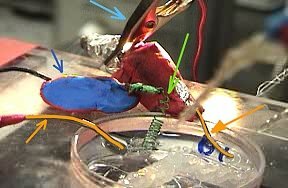 In the dish are: perfusion tubes (colored orange and marked with orange arrows) to exchange the solution in part of the dish; the ground wire (colored green and marked with green arrow). The ground wire is affixed to the microscope stage with some children's modeling clay (colored blue). The alligator clip (light blue arrow) grounds the perfusion tubing and the bath. |
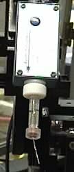 Here's the headstage (a small preamplifier) with the electrode holder attached. Here's the headstage (a small preamplifier) with the electrode holder attached. |
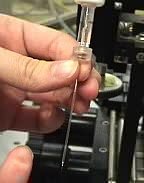
 This is the intracellular pipette. It can be used to stimulate AND record. It is usually filled with KCl and KAC. This is the intracellular pipette. It can be used to stimulate AND record. It is usually filled with KCl and KAC. |
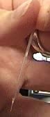
 The glass pipette fits over the wire that comes out of the electrode holder. The solution in the glass pipette must contact the wire of the electrode holder. The glass pipette fits over the wire that comes out of the electrode holder. The solution in the glass pipette must contact the wire of the electrode holder. |
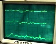
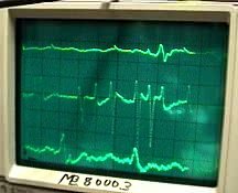 Representative voltage recordings of spontaneous activity from three of the 4 electrodes. |
 |
| Overview | Web Lectures | Fellowships | Activities | Home | SFN | NAS | IBRO |