 Return to Methods List |
Methods in Neuroscience Manduca sexta Segmental Ganglia - Dissection p. 2 |
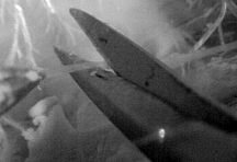
| Below are photos of what the ganglia look like in situ. To dissect out the nerve cord (or ganglion), muscles, trachea and fatty tissue must be cut or removed. Neither the photos below nor the video illustrate the complete dissection. However, the video will give you an idea of the slow speed at which the dissection is done and of how the tissues that need to be removed are handled. Also from the video you should be able to see that removing the nerve cord is not difficult. The nerve cord is easy to visualize; you just need to be slow and careful when removing it. |
|
Dissecting out the nerve cord Click HERE for a 1 minute video (10MB) |
|
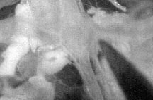
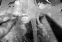
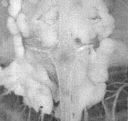 Here is one ganglion sitting on top of some fatty and muscle tissues. |
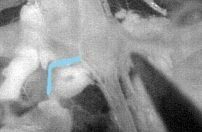
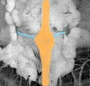 Here the web designer colored the ganglion orange and the easily visible nerve roots blue. |
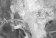
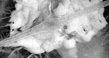 Another ganglion - more nerve roots are visible. |
;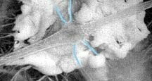 Nerve roots colored blue. |
 Muscles, trachea and fatty tissue must be cut or removed to visualize the nerve cord (or ganglion). |
 |
 The nerve roots on each ganglion must be cut. |
 Here the nerve root is colored blue. |
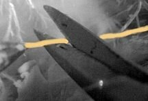
 Here the forceps are holding the nerve root. |
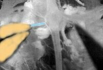 Forceps are colored orange. |
 One ganglion after the nerve roots have been cut. |
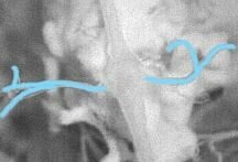 Nerve roots are colored blue. |
| Overview | Web Lectures | Fellowships | Activities | Home | SFN | NAS | IBRO |