 Return to Methods List |
Methods in Neuroscience Manduca sexta Segmental Ganglia - Recording p. 1 |
| Below are photos showing how the ganglia are pinned in the recording dish. |
|
How the nerve cord is pinned in the recording dish Click HERE for a one minute video (10MB) |
|
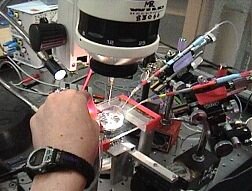 The microscope stage: in the middle you can see the dish in which the ganglia are pinned. The 3 suction recording electrodes come in from the right. The intracellular recording electrode is located at the far left and not visible in this photo. |
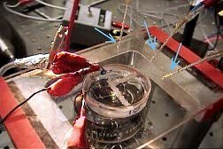 In the center of the photo is the dish containing the ganglia. Some of the ganglia are isolated by a thick "wall" of petroleum jelly. In the dish are also a ground wire and a perfusion system that circulates the bath solution in part of the dish. A close-up photo of the dish contents is shown on the recording 3 page. The ends of the three suction recording electrodes are marked with blue arrows. |
 The six ganglia are shown by the orange arrows. From the left, going counterclockwise, they are ganglia 2,3,4,5,6 and the fused ganglia #7-11. |
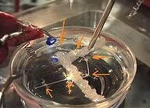
 Here the web designer has colored the wall of petroleum jelly orange. |
 Here the web designer has colored the two different, solutions yellow and blue. The perfusion system refreshes the solution that is colored yellow. |
|
|
Close-up view of all the ganglia Click HERE for 15 second video (2.6MB) |
|
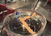
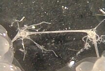 A close-up view of ganglia 5 and 4 (from left to right). |
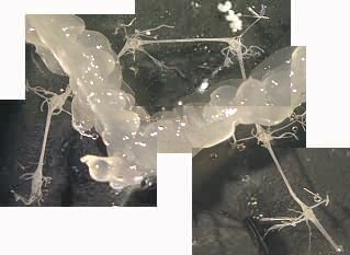 A close up view of the entire nerve cord. Clockwise from the left: the fused ganglia numbers 7-11, then ganglion 6, 5, 4, 3 and 2. |
| Overview | Web Lectures | Fellowships | Activities | Home | SFN | NAS | IBRO |