 Return to Methods List |
Methods in Neuroscience Scallop Retina - MAIN PAGE |
|
All the photos here were taken at the Marine Biological Laboratory in Woods Hole, Massachusetts during the summer of 2001. Many thanks to Enrico Nasi and especially to Maria Gomez for their enthusiasm and willingness to spend time explaining their work. Please note that most of the photos on these web pages were taken either in the dark, or under dim, red light. This is why the scientists' photos, below, are strange colors. |
||
MEET THE SCIENTISTS Maria del Pilar Gomez, MD, PhD Research Assistant Professor, Dept. Physiology & Biophysics, Boston University |
MEET THE SCIENTISTS Enrico Nasi, PhD Professor, Dept. Physiology & Biophysics, Boston University |
MEET THE SCIENTISTS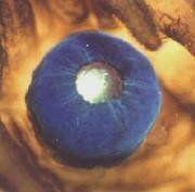 Meet the eye of the Bay Scallop, Pecten irradians |
| Overview |
|
In the web pages shown here, Maria Gomez and Enrico Nasi have taken advantage of the unusual eyes of marine invertebrates, such as Pecten irradians. These eyes are highly developed, strikingly resemble the gross morphology of their vertebrate counterparts, and present the remarkable feature of a double retina, in which both depolarizing and hyperpolarizing responses to light had been previously reported. Nasi and Gomez developed a preparation of enzymatically dissociated photoreceptors which remain physiologically viable. The major benefits of this approach include the elimination of all confounding cell-cell interactions, the unambiguous morphological identification of cell types, and the removal of all surrounding connective tissue. Removal of the connective tissus completely exposes a clean surface of the plasma membrane, making the cells ideal for electrophysiological recording via patch micropipettes. They used the whole-cell patch clamp technique to determine that in solitary cells presenting a microvilli-covered lobe (called the rhabdomere, and similar to the light-sensing structure of other invertebrate photoreceptors) light evokes an inward current. By contrast, in other photoreceptor cells - which are characterized by the presence of ciliary appendages (which are structurally analogous to the light-transducing outer segment of vertebrate rods and cones) - an outward photocurrent was recorded. Because the cells are isolated and synaptic interactions therefore can be ruled out, these results conclusively established the coexistence of two fundamentally distinct classes of photoreceptors in these retinas. These distinct classes represent the two known lineages of light-sensing cells found across species. This exceptional model system provides an opportunity to investigate the divergence in the signalling pathway that couples photon absorption to the generation of an electrical response. |
| Overview | Web Lectures | Fellowships | Activities | Home | SFN | NAS | IBRO |