 Return to Methods List |
Methods in Neuroscience Scallop Retina - DISSECTION 1 |
|
Some of the key steps in the initial dissection of the scallop eye Click HERE for a 30-second (4.7MB) video showing this. |
|
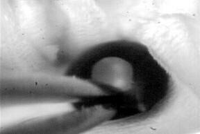 A small piece of the "pigmented layer" (the equivalent of our sclera) is grasped with the forceps... |
 and the scissors are used to cut off this small piece of pigmented layer. |
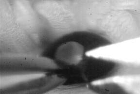
 scissors cutting... |
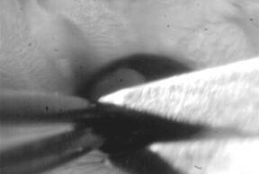
 scissors finishing cutting. |
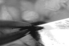
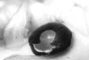 This is what the eye looks like after the small section has been cut off. |
 Next, the pigmented layer on one side of the cut is held with the forceps and the scissors are used to cut around the eye. |
 From this photo you should be able to get a good idea of how much of the pigmented layer is going to be removed. |
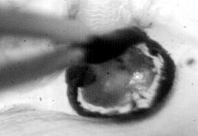
 Here is what the eye looks like as the cutting around the eye begins. |
 Cutting around the eye continues until a ring has been cut off from all around the eye... |
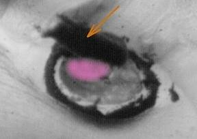 and the piece of pigmented layer (orange arrow) can be removed. This leaves the bottom half of the eye, on which the retina lies. Go on to "dissection 2" to see how the retina is removed. The object the website designer has colored pink is the lens. This must be lifted out before the retina can be removed. |
| Overview | Web Lectures | Fellowships | Activities | Home | SFN | NAS | IBRO |