 Return to Methods List |
Methods in Neuroscience Scallop Retina - DISSECTION 2 |
|
Some of the key steps in the removal of the scallop retina Click HERE for a 15-second (2.1 MB) video showing this. |
|
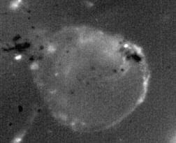
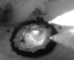 The retina resides on the bottom side of the scallop eye. You can see that here the edge of it is loose. Once the pigmented tissue is dissected out, the retina is removed from the back of the eye by cutting the edges which attach it to the optic nerves. Here it looks very easy to remove the retina, but in reality it takes some practice. |
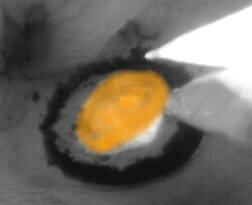 Here the website designer has colored the retina orange so you can see it easier. The tip of the scissors is being used to gently lift up the retina. |
 The orange arrow points to the edge of the retina. |
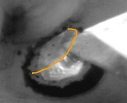 Here the edge of the retina is outlined in orange. The retina is basically transparent. |
 The retina is gently lifted off the eye... |
 into the artificial seawater bathing solution. |
 Here the retina is out of the eye and in a pronase solution (an enzyme treatment). After pronase treatment, the cells are mechanically dissociated. |
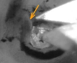
 After the scallop eye has been damaged, you can see a tentacle getting wrapped over the eye in an attempt to protect it. |
| Overview | Web Lectures | Fellowships | Activities | Home | SFN | NAS | IBRO |