 Return to Methods List |
Methods in Neuroscience Squid Giant Synapse - MAIN PAGE |
| All the photos here were taken at the Marine Biological Laboratory in Woods Hole, Massachusetts during the summer 2001 Neurobiology course. Many thanks to the course instructors and students who made these pages possible. | ||
 |
SQUID TEAM Pam Follett, neurobiology course student FELIX E. SCHWEIZER, PhD, Assistant Professor of Neurobiology, UCLA, neurobiology course instructor Ernie Barreto, neurobiology course student |
 |
| OVERVIEW |
|
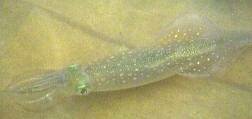
The squid giant synapse is one of the classical preparations for studying synaptic physiology. The synapse can be dissected with relative ease and in a good specimen synaptic transmission can stay functional for many, many hours.
The giant synapse is located in the stellate ganglion and is part of the giant fiber system, which likely is involved in the escape response of squid. The giant fiber system is made up of three consecutive sets of axons, each of which results from the fusion of many smaller axons. The first order giant axon sends information received from sensory cells to the second order axon which leaves the brain and enters the stellate ganglion. There, the second order giant axon fans out like a hand and each finger contacts one of the giant third order axons that exit the ganglion and innervate the musculature of the mantel. The third order axons that innervate portions of the mantel close to the neck are relatively small, while the one that innervates the tail end of the squid is by far the largest in diameter. It is this largest giant axon (THE giant axon) that was used by Hodgkin and Huxley for their famous experiments. The largest synapse, the one made onto THE giant axon, is the one which is routinely used for studies of synaptic transmission as it offers best access to both pre- and postsynaptic elements of the synapse. The squid giant synapse contains hundreds of release sites, much like the neuromuscular junction or the more recently popular Calyx of Held in the mammalian auditory system. Synaptic vesicles cluster at each of these release sites and each site is contacted by a projection from the postsynaptic axon. In the isolated giant synapse preparation, many different recording configurations are possible. An electrode inserted into the second order axon before it enters the stellate ganglion is frequently used to trigger action potentials. This action potential then invades the presynaptic nerve terminal where it can be measured by a second electrode. A third electrode inserted into the postsynaptic cell measures the postsynaptic response which gives a good report of the amount of neurotransmitter release. Both, the presynaptic and the postsynaptic terminals can also be voltage clamped by inserting additional electrodes. The electrodes can also be used to inject reagents thought to disrupt synaptic transmission. If injection is stopped the reagents will dilute into the large pre- or postsynaptic intracellular volume and reagent induced effects can thus reverse. This is only one of the many unique advantages of this preparation. Selected additional reading: George J. Augustine, Marie E. Burns, William M. DeBello, Sabine Hilfiker, Jennifer Morgan, Felix E. Schweizer, Hiroshi Tokumaru and Keiko Umayahara. (1999). Proteins Involved in Synaptic Vesicle Trafficking. Journal of Physiology 520, 33-41. Rodolfo R. Llinas (1999) The Squid Giant Synapse : A Model for Chemical Transmission, Oxford University Press; ISBN: 0195116526. |
| Overview | Web Lectures | Fellowships | Activities | Home | SFN | NAS | IBRO |