 Return to Methods List |
Methods in Neuroscience Squid Giant Synapse - DISSECTION - Page 1 |
| Below are photos of some of the key steps in the initial dissection of the squid to expose the stellate ganglion. |
|
Dissection - first steps Click HERE for a 4.5 min video (42MB) |
||
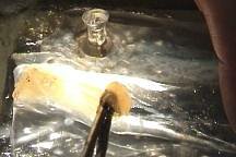
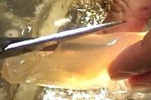 After the head was removed, a long cut is made ventrally in the anterior to posterior direction... |
 and the squid is then laid flat and pinned down. |
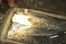
 Some internal organs of the squid can be observed: ink sack (green), egg maturation organ filled with red symbiotic bacteria (blue), gills (lavendar), egg sack (pink). |
 The stellate ganglion is visible beneath the muscle that surrounds the hepatopancreas. |
 - a more close up view of the branches of the ganglion (indicated by the forceps) |
 The gills are removed bilaterally. |
 The ink sack is removed by pulling posteriorly. |
 The siphon, with which squid swim, is opened. |
 The siphon muscle is cut away, being careful because the stellate ganglion is just below it (not visible in this photo). |
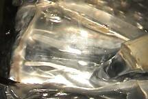
 The digestive system (the hepatopancreas) is opened. It is basically a hollow tube. |
 The hepatopancreas is grabbed from the back and removed by pulling it anteriorly (left in the photo). |
 Wash with sea water to get rid of all the enzymes from the hepatopancreas. |
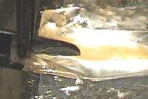
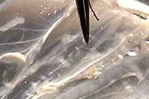
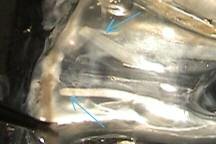 Two presynaptic axon bundles can be seen (blue arrows). They are running inside the hepatopancreas. |
 The axon bundle is tied off (only on the side from which recordings will be made). |
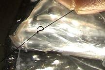 The string is pulled tight. This serves to tie off cut axon ends, but the string is also handy for holding onto and manipulating the preparation. |
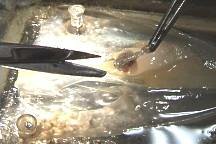
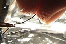
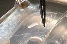
 At this stage many aspects of the nervous system can be observed, such as the nerve fibers that connect both sides of the giant fiber system (where the forceps are pointing). |
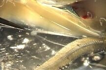
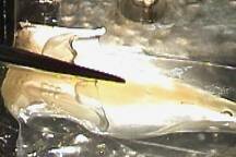
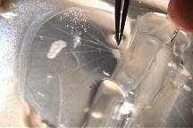
 Here you can see the stellate nerves emanating from the stellate ganglion (forceps point to one stellate nerve). Each nerve fiber contains one giant axon, plus many smaller axons. The giant axon and most of the smaller ones innervate the mantel musculature. The largest giant axon (the one used by Hodgkin and Huxley) is running parallel to the animal's axis and is obscured in this picutre. |
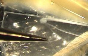
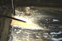
 The stellate ganglion is located here (forceps are pointing to it), just under the muscle that encloses the hepatopancreas. 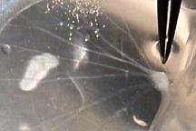 |
| Overview | Web Lectures | Fellowships | Activities | Home | SFN | NAS | IBRO |