 Return to Methods List |
METHODS in Neuroscience Whole Cell Patch Clamp Technique - MAIN PAGE |
| The photos here were taken at the Marine Biological Laboratory in Woods Hole, Massachusetts during the summer 2001 Neurobiology course. Many thanks to the course instructors and students who made these pages possible. | |
 WHOLE CELL TEAM WHOLE CELL TEAMKAMRAN KHODAKHAH, PhD, Assistant Professor of Neuroscience at Albert Einstein College of Medicine, neurobiology course instructor Tobias Rasse, neurobiology course student Shelly Emond, neurobiology course student |
 |
| OVERVIEW |
|
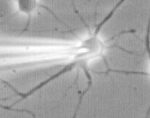
The technique of patch clamp recording was invented by Bert Sakmann and Erwin Neher in 1981 for which they received the NOBEL prize. Without doubt, the patch clamp techniques is one of the most powerful techniques in the study of physiology, and one that has revolutionized the field. The technique is best suited for the study of the behaviour of single ion channels, or macroscopic currents in small cells or macro-patches. The whole cell technique allows one to control the composition of solutes on both sides of the membrane. This is an invaluable tool in determining the biophysical properties of currents under study. The techniques is easily combined with other techniques such as fluorescence microscopy and flash photolysis. In recent years, for example, the whole cell patch pipette has been used to introduce into the cell ion selective fluorophores for quantitative measuremnts of calcium, magnesium, sodium and proton concentrations. Similarly, various caged compounds have been introduced into the cell to study the role of second messengers in the cell by flash photolysis.
|
| Cell culture (Courtesy of Dr. Jeanne M. Nerbonne, Professor of Molecular Biology and Pharmacology, Washington Univ. School of Medicine, St. Louis, Missouri, USA) |
||
 Patch clamp experiments often use cultured cells. Here Amy in the Nerbonne lab prepares cells under sterile conditions in the hood. |
 Mamallian cells are kept in an incubator, often in small culture dishes. The media is pink due to the pH indicator. |
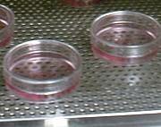 A close up view of the culture dishes in the incubator. |
| Overview | Web Lectures | Fellowships | Activities | Home | SFN | NAS | IBRO |