 Return to Methods List |
Methods in Neuroscience in vitro Slice Preparation - RECORDING EQUIPMENT |
| Below are photos of some of the equipment used to make in vitro slice recordings. The main requirements for sucessful visualized slice recording are mechanically stable recording chambers and micromanipulators, a microscope with a focusing nosepiece (where the objective moves, rather than the stage) that sits on a translation (moveable) stage, water-immersion objectives, and the same amplifiers, etc. used for other types of patch clamp recording. It should also be noted that some types of electrophysiological recordings can be made from brain slices using far simpler equipment than shown here. |
| Recording equipment | ||
 Microscope with differential interference contrast (DIC) optics. The recording electrode can be seen on the manipulator at the right. |
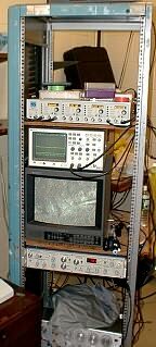 |
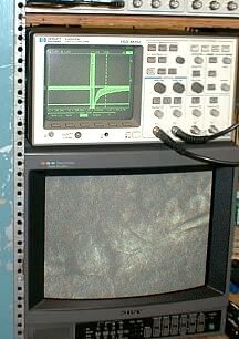 Since the slices are illuminated with near-infrared light, which is out of the range of human vision, a video monitor is required to see what's going on. It is also a useful teaching tool. Here the oscilloscope shows currents being recorded from the neuron. |
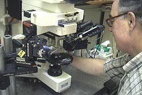 Here Joe is carefully lowering the glass pipette into the recording chamber in much the same way as is done in whole cell or single channel recording. |
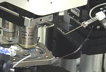 A close up view of the microscope stage and the glass recording pipette. |
| Overview | Web Lectures | Fellowships | Activities | Home | SFN | NAS | IBRO |